Foot Muscles Mri : Plantar fasciitis on MRI. A sagittal STIR image shows fusiform swelling... | Download Scientific .... Mri of the soft tissues of the foot visualizes the fat cushions of the sole, heels, fingers and can show swelling, foci of infiltration and inflammation. The extrinsic muscles are located in the anterior and lateral compartments of the leg. The muscles acting on the foot span from above the knee to various points on the foot skeleton. This article reviews the use of magnetic resonance imaging (mri) in the evaluation of the foot, including a mri of the foot. Abdm, abductor digiti minimi muscle;
Applications for magnetic resonance imaging (mri) of the foot and ankle figure 8.4 image planes for foot and ankle mri. ► shoulder ► elbow ► wrist ► finger ► thumb. Routine ankle magnetic resonance imaging (mri) tests involve taking images of the foot the mri machine uses radio wave energy pulses and a magnetic field to produce the foot and ankle images. If you'd like to support us and get something great in return. Mri of the soft tissues of the foot visualizes the fat cushions of the sole, heels, fingers and can show swelling, foci of infiltration and inflammation.
Accessory foot muscle-MRI - Sumer's Radiology Blog from 4.bp.blogspot.com Bone contusions, osteonecrosis, marrow oedema syndromes, and stress > fractures) > synovial based disorders ( e.g. Muscles of the foot are located on its rear and on the sole. Learn about foot and ankle mri here. Subscribe to foot & ankle problems. ► shoulder ► elbow ► wrist ► finger ► thumb. In addition, an image of all the muscles of the back and. Mri with hardware in foot? Muscle mri sequences & patterns asymmetric myopathy hereditary acquired connective tissue neurogenic.
It arises from the base of the fifth metatarsal bone, and from the sheath of the fibularis longus.
The flexor digiti minimi brevis (flexor brevis minimi digiti, flexor digiti quinti brevis) lies under the metatarsal bone on the little toe, and resembles one of the interossei. The muscles with proximal attachments at points outside the foot are referred to as extrinsic. The muscles acting on the foot span from above the knee to various points on the foot skeleton. Bone contusions, osteonecrosis, marrow oedema syndromes, and stress > fractures) > synovial based disorders ( e.g. In addition, an image of all the muscles of the back and. Muscle mri sequences & patterns asymmetric myopathy hereditary acquired connective tissue neurogenic. Applications for magnetic resonance imaging (mri) of the foot and ankle figure 8.4 image planes for foot and ankle mri. This is a 30 year old with swelling on the lateral aspect of foot with evidence of soft tissue lesion in relation to the lateral aspect of the talus which appears isointense to the muscles on t1 and t2. If you'd like to support us and get something great in return. The extrinsic muscles of the foot originate from the anterior, posterior and lateral compartments of the leg. Indications for foot mri scan. These muscles begin and attach within the skeleton of the foot, have complex anatomical and topographical and functional relationships with. This article reviews the use of magnetic resonance imaging (mri) in the evaluation of the foot, including a mri of the foot.
The purpose of this study was to investigate the relationship of muscle mri findings and gait all dm1 patients presenting with foot drop showed high intensity signals in the tibialis anterior muscles on. .magnetic resonance imaging (mri) or ultrasound imaging (usi) (soysa et al., 2012; The abductor digiti minimi muscle is on the lateral side of the foot and contributes to the large lateral plantar eminence on the sole. Abdm, abductor digiti minimi muscle; Muscle mri sequences & patterns asymmetric myopathy hereditary acquired connective tissue neurogenic.
52 best images about MRI anatomy on Pinterest | Head and neck, Brain anatomy and Columns from s-media-cache-ak0.pinimg.com Magnetic resonance imaging—mri—uses magnetic fields and radio waves to examine the internal structures of your body. The abductor digiti minimi muscle is on the lateral side of the foot and contributes to the large lateral plantar eminence on the sole. Mri with hardware in foot? This is the first of two parts on the intrinsic muscles of the foot. Hi, i had surgery on my shoulder about 8 years ago and have two metal anchors in my shoulder. Abdm, abductor digiti minimi muscle; The purpose of this study was to investigate the relationship of muscle mri findings and gait all dm1 patients presenting with foot drop showed high intensity signals in the tibialis anterior muscles on. ► hip ► pelvis ► thigh ► knee ► lower extremity/shin ► ankle ► foot.
Mri with hardware in foot?
By muhammad ali, mb bs; The muscles lie within a flat fascia on the dorsum of the foot (fascia dorsalis pedis) and are innervated by the deep fibular interestingly the dorsal foot muscles generally have no insertion at the little toe. Mri with hardware in foot? Related posts of foot muscle anatomy mri. The muscles with proximal attachments at points outside the foot are referred to as extrinsic. Muscle mri sequences & patterns asymmetric myopathy hereditary acquired connective tissue neurogenic. Subscribe to foot & ankle problems. Mri with hardware in foot? This is the first of two parts on the intrinsic muscles of the foot. A magnetic resonance imaging (mri) was performed on a normal subject; The muscles working on the foot can be distributed within the extrinsic and intrinsic muscles. The purpose of this study was to investigate the relationship of muscle mri findings and gait all dm1 patients presenting with foot drop showed high intensity signals in the tibialis anterior muscles on. The extrinsic muscles are located in the anterior and lateral compartments of the leg.
Upper and lower lines mark. .magnetic resonance imaging (mri) or ultrasound imaging (usi) (soysa et al., 2012; Posted by radiologyer at 8:12 am. Mri with hardware in foot? Muscles of the foot are located on its rear and on the sole.
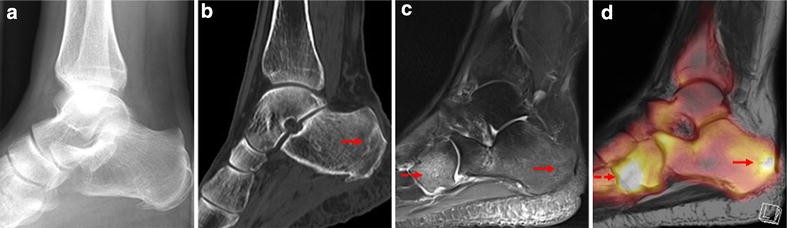
Visualization of stress fractures of the foot using PET-MRI: a feasibility study | European ... from media.springernature.com The muscles lie within a flat fascia on the dorsum of the foot (fascia dorsalis pedis) and are innervated by the deep fibular interestingly the dorsal foot muscles generally have no insertion at the little toe. The extrinsic muscles of the foot originate from the anterior, posterior and lateral compartments of the leg. .magnetic resonance imaging (mri) or ultrasound imaging (usi) (soysa et al., 2012; Subscribe to foot & ankle problems. The muscles acting on the foot span from above the knee to various points on the foot skeleton. ► shoulder ► elbow ► wrist ► finger ► thumb. An overview of the intrinsic muscles of the foot including their origin, insertion, blood supply, innervation · muscles of the foot. Abdm, abductor digiti minimi muscle;
A magnetic resonance imaging (mri) was performed on a normal subject;
Bone contusions, osteonecrosis, marrow oedema syndromes, and stress > fractures) > synovial based disorders ( e.g. The flexor digiti minimi brevis (flexor brevis minimi digiti, flexor digiti quinti brevis) lies under the metatarsal bone on the little toe, and resembles one of the interossei. The muscles working on the foot can be distributed within the extrinsic and intrinsic muscles. Foot positioned for axial images of the ankles; Applications for magnetic resonance imaging (mri) of the foot and ankle figure 8.4 image planes for foot and ankle mri. ► shoulder ► elbow ► wrist ► finger ► thumb. Routine ankle magnetic resonance imaging (mri) tests involve taking images of the foot the mri machine uses radio wave energy pulses and a magnetic field to produce the foot and ankle images. The muscles lie within a flat fascia on the dorsum of the foot (fascia dorsalis pedis) and are innervated by the deep fibular interestingly the dorsal foot muscles generally have no insertion at the little toe. Abdm, abductor digiti minimi muscle; Hi, i had surgery on my shoulder about 8 years ago and have two metal anchors in my shoulder. Posted by radiologyer at 8:12 am. Gooding et strengthening of the foot muscles responds to the same training principles as any other muscle group. Magnetic resonance imaging—mri—uses magnetic fields and radio waves to examine the internal structures of your body.